The Wrong Punch Line

Patients will often withhold information from their medical providers for numerous reasons. It could either be due to the embarrassment, the idea that it may not be important, the fear of getting into trouble, or protecting someone else. There were reasons all across the board. It can be quite difficult for medical providers to properly treat a patient if patients do not inform providers of all the circumstances. Medical providers are not there to judge the patient. They are not there to scold patients for their actions. They are here to make sure that their patients are alive and okay. Tattling does not come with our job titles.


But as a 13 year old in middle school, I can see why medical professionals seem scary -especially in front of parents.

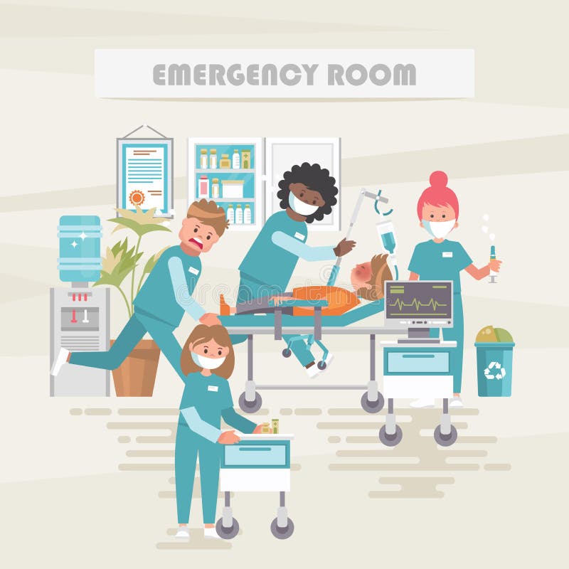
It was another afternoon in the ER when we met our 13 year old male patient who was accompanied by his mother for abdominal pain. More specifically, the pain was regional to the left-sided flank area with no associated symptoms. The initial suspicion for flank pain is usually kidney stones. Although kidney stones are more common in adults (for numerous reasons such as certain prescription medications or dehydration), it is not rare for teenagers. The patient, a fairly skinny boy, looked distressed. He was squirming in his chair and relied on his mother to speak with the doctors. I can’t judge him though. I used to be a shy kid too. 


Our patient was having a regular day at school when he started feeling pain near his right hip. The pain was consistent, non-radiating, and was not relieved by any factors. The mother was concerned because the patient woke up completely fine and went to school without any complaints. She gave her son Pepto Bismal when he came home from school as they suspected he had an upset stomach from the school lunch. The thought of having to consume school lunch made even my stomach turn. Unfortunately, the pain persisted, which prompted the mother to bring her son to the ER for further evaluation. 


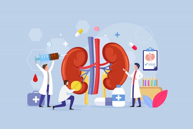
Indicators of kidney stones are typically severe abdominal pain, nausea, vomiting, and dysuria (pain with urination). Many individuals may also experience hematuria (blood in the urine). Abdominal pain can be caused for numerous reasons. The best way to determine the etiology of the abdominal pain, or any medical concern for that matter, is first through the history obtained from the patient. In this case, there were no outside factors that were contributing to this patient’s abdominal pain. The patient did not consume any takeout or new food this past week to suggest an upset stomach. The lack of fever, nausea, and vomiting ruled out a possible bacterial infection. There were also no recent injuries that could account for the abdominal pain. Considering all viewpoints, the next test would be a urinalysis. A urine sample can result in multiple findings: for kidney-related issues, physicians will look for red blood cells that indicated hematuria, crystals, and trace minerals that could lead to stone formation, white blood cell elevation to account for infections (especially urinary tract infections) and also increases in the urine pH which can also indicate kidney stones. It is not necessary for all of those elements to come back positive to suggest kidney stone formation, but it will give the physician enough evidence to start treating the condition properly and effectively. To our surprise, the urinalysis was completely normal. 

The First Signs of Kidney Stones: Pain, Causes, Treatment & Passing


So what was going on? 

Pain medication that was supposed to alleviate the abdominal pain was not working. In fact, the patient was complaining of increasing abdominal pain which was now associated with dysuria. Things were just not adding up. No infection. No stones. No food poisoning. And certainly no indication for discharge. This meant further testing. 

The mother at this point was frustrated with the 2-hour duration of the ER visit. She wanted to go home and see if her son could just sleep it off. The patient looked tired too. But the risk of missing something significant was not worth it. The physician decided to obtain a CT scan of the abdomen for further investigation. Perhaps there were small salt deposits that were in the kidneys that were not picked up by the labs, but could potentially be seen through a scan. 

We got the scans back and it was a finding that the physician would not have considered given the initial history. He asked the mother if it was okay to step outside of the room so that he could speak to the patient in private. 

Doctor: “Hey, how are you feeling?”

Patient: “Not so good. Nothing changed”.

Doctor: “I’m sorry to hear that. Is there anything you would like to share with me that you didn’t feel comfortable sharing in front of your mom?

The patient hesitated for a minute. He started to shake his head when the physician interrupted. 

Doctor: “You’re not gonna get in trouble, that’s my promise to you. But I can’t help you feel better if I don’t know the full story”

Patient: “We were in gym class and I was joking around with my friends and this girl accidentally punched me.”

Doctor: “That must have been one strong accidental punch. Can I invite your mother back in?”

The boy looked down and nodded. 

“Your son’s left kidney appears dented on the scan. This is usually the result of a blunt force trauma.”

CT scan of a kidney with blunt force trauma


The mother looked confused- rightfully so. The patient was not in a car accident, nor was he injured by any blunt force object. 

“He actually got punched, accidentally, on the left side of his abdomen. Due to his small frame and low weight he was punched hard enough to sustain an injury to the kidney. We’re going to transfer him to the pediatric ER for further management.”

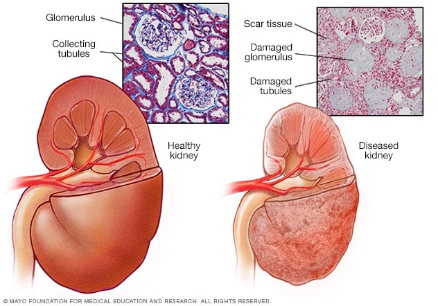
The mother looked horrified and confused. Although the patient wasn’t at any immediate risk, it was important to monitor his kidneys to prevent any future renal complications which could include dysuria, hematuria, infection, etc. Most cases, like this, will be treated non-operatively. Non-invasive treatments such as checking vitals, urine cultures, and lab work that assesses the function of the kidneys are necessary to clear the patient for discharge. Bed rest is also crucial in making sure the body can recover from the added stress. 

While it was a long ER visit and an even longer story, it seemed like our patient’s friend got the punchline wrong. 

Sources:
https://jamanetwork.com/journals/jamapediatrics/fullarticle/2442837
https://www.niddk.nih.gov/health-information/urologic-diseases/kidney-stones/diagnosis
https://www.ncbi.nlm.nih.gov/pmc/articles/PMC6120183/

Update

Hello Readers,

So…it’s been some time. Approximately 8 months, but who’s counting, right? I would say it took me all this time to come up with the title for my next post, which isn’t entirely false. To be quite honest, I took some time off to focus on some things, but then I didn’t figure out how to come back. I’d like to blame some of it on writer’s block and the rest on lack of motivation.


That said, I really do miss writing up these stories and doing my research on interesting case studies. I have a few blog posts in the works, and so many more I’m excited to write. I’m hoping to finalize these soon and have it posted.

Hmm, I did get a cat back in November


Thank you for your continued (and shared) interest in these stories and being patient with me! I don’t think I will ever be able to describe the joy I get from being able to share these stories with you.

Here’s to more Med Thoughts 🙂


Hang in there Ryerson, and we'll see you soon - The Eyeopener

Saturday Night Live

There is absolutely no way to know what to expect when you walk into an emergency room. There can be a trend of kidney stone patients, children with flu-like symptoms, and if it’s a weekend most likely a stream of EtOH intoxication patients. But this night… this was an interesting night.

It was surprisingly a slow Saturday night at the ER. I know what you’re thinking, did I get the day of the week wrong? A quiet Saturday? That happens? It’s rare and when it happens, absolutely no one is allowed to mention it. Rule number one of working at a hospital: never ever mention how “quiet” or “slow” the floor is because you will regret it. It’s almost like the universe hears us cheering the very rare downtime and blesses us with a board full of all the complicated patients. Trust me, we’ve all done it and then regretted it immediately. It’s nice to have a breather and take a seat after running around patient after patient.

It was around 4AM when we got the notification. A young male enroute s/p trauma. Now, we are not a designated trauma ER and often times the critically traumatic cases are routed to the nearest trauma units. In this case, the closest hospital to the patient was our hospital and the patient needed the immediate medical attention for not just one, but three gunshot wounds.

44,760 Ambulance Staff Illustrations, Royalty-Free Vector Graphics & Clip  Art - iStock

I walked into the chaos of the critical care room and it is swamped with nurses ready with their crash cart necessities, the attending provider, the PAs and med techs. There was also a few other medical staff nearby to tune in; all of our minds curious about what was happening. We so rarely witnessed gunshot wound patients that when one does make his way to triage, everyone was interested.

In came the patient on a stretcher and curled up in distress. Our lovely medical staff were not the only ones witnessing his moans and groans .. he was streaming live on social media while being transported from the stretcher to hospital bed. “Hey MTV, welcome to my deathbed?” I guess you can to do anything for the most engaging content. I would recommend a trigger warning next time.

There was an actively bleeding wound, covered with gauze on the left side of his neck and his left shoulder blade. His pants were ripped and stained with blood all over his back side. every single medical personnel was busy doing their specific task. All the noise is almost harmonious and can feel like there is a ticking clock right inside of your heart. The nurses were quick with no time to waste with their crash cart ready, IV line in place, while the med techs worked in synch to put on the EKG nodes and switching the patient into the very fashionable blue hospital gown.

Emergency Room Stock Illustrations – 6,888 Emergency Room Stock  Illustrations, Vectors & Clipart - Dreamstime

Then, one of my favorite no b.s. nurses ever so sweetly shouted, “Sir…this is NOT the time or place to be live-streaming. Put that damn phone away”. It was pretty nice to see a phone flying across the critical care room. You don’t really get to see that happening on a day to day basis.

The first ten minutes, “platinum minutes”, after a person is shot is absolutely critical to make sure all the right protocols are followed. The bleeding needs to be identified and stopped as much as possible. Applying pressure to the wound can prevent the patient from excess bleeding which can lead to hemodynamic shock. Gunshot wounds to the body cavities/trunk pose greater risks due to hemorrhaging and organ injury. Although the entrance wound can be addressed, there is no way to decrease the internal injuries without the proper medical attention (usually emergency surgery). The earlier, the better as it can prevent ischemia, organ failure, and death. Extremity gunshot wounds are a little easier to address as the bleeding can be temporarily stabilized with a tourniquet. Worse case scenario would be limb ischemia which is still less riskier than organ injury.

An external file that holds a picture, illustration, etc.
Object name is Dtsch_Arztebl_Int-114-0237_001.jpg

Before a patient is considered for surgery, medical providers will do their best to manage the wound and stop any further injuries from occurring. Surgery can pose a risk as there can be organ dysfunction which can further complicate the surgery and healing process. When a gunshot wound patient arrives at the ER, the medical providers make sure the patient is getting transfusions if there was too much blood loss, pain control, coagulation to increase blood coagulability, airway protection if needed, and monitoring of vitals at all times. To determine the location of a bullet that is lodged into the body, the patient will need a CT scan. This scan will provide valuable information of not only the location of the foreign object, but also the level of internal damage. Bullet wounds in the body cavities are dangerous because it can lead to organ perforation (essentially, a hole in the organ). Even if the bullet did not directly strike the organ, these are post-trauma side effects. Bowel perforation is the most common after a gunshot wound. Many patients return to the ER many months and even many years later with small bowel obstruction due to a prior gunshot wound.

Three bullet wounds may seem like a death sentence. Our live-streaming patient was on the luckier side. Two of the three bullet wounds were just a result of brush fire (the bullets slid past the patient causing abrasions without actually entering the body). The third bullet though, that one was a problem. Unfortunately for the patient, the third bullet was shot at his rear and went into his colon. Gunshot wounds in the colon are more sensitive to possible infections during the healing process. In order to prevent cross contamination from the large intestine while wound healing, surgeons will perform a colostomy. A colostomy is a surgical procedure in which one end of the large intestine is surgically placed outside of the abdominal wall within a stoma (opening in the skin where the colostomy bag is placed). Stool is collected in the colostomy bag which is cleaned out manually in order to provide the gunshot wound the space to heal.

Ostomy

Our live-streaming patient was then transferred to another facility for further medical management. He was alive, stable, and extremely lucky. After that notification, the ER pace slowed down a bit as it gradually does around 4:30AM. We were sitting around, yawning, stretching, and also processing the situation.

One of the medtechs popped in shortly after trying to contain his laughter. “Y’all made it onto the live,” he says laughing and pulling up the patient’s Instagram account.

There we were on the phone screen for the briefest minute, scrubs, gloves, and intensively bright lights, doing what we did best on Saturday Night Live.

References:

Cimino-Fiallos, Nicole. “Gunshot Wounds: A Targeted Approach.” Medscape Drugs & Diseases – Comprehensive Peer-Reviewed Medical Condition, Surgery, and Clinical Procedure Articles with Symptoms, Diagnosis, Staging, Treatment, Drugs and Medications, Prognosis, Follow-up, and Pictures, 28 Jan. 2019, reference.medscape.com/slideshow/gunshot-wounds-6008960.

Drummond, Hamilton. “Gunshot Wounds of the Large Intestine and Rectum, with Special Reference to Surgical Treatment.” Proceedings of the Royal Society of Medicine, vol. 13, no. Surg_Sect, 1920, pp. 24–34., doi:10.1177/003591572001302117. “Home – PMC – NCBI.”

 National Center for Biotechnology Information, U.S. National Library of Medicine, http://www.ncbi.nlm.nih.gov/pmc/. 

Webster, J. E., and E. S. Gurdjian. “Acute Physiological Effects Of Gunshot And Other Penetrating Wounds Of The Brain.” Journal of Neurophysiology, vol. 6, no. 4, 1943, pp. 255–262., doi:10.1152/jn.1943.6.4.255. 

Kidney Drama and Dialysis

After some observation, I’ve come to the conclusion that the kidneys are in fact the most dramatic organs. Your body could be dealing with an issue such as a pulmonary embolism and your kidneys will somehow manage to steal the spotlight. These are all just observations though…I’ll have to do some further investigating before I can add more dirt for those kidneys to filter out. It just goes to show how the human body functions as a whole system, rather than separate systems.

Kidneys have a lot on their plate. The kidneys balance the body by filtering out excess fluid, removing waste, regulating blood pressure, and monitoring red blood cell production. Unfortunately, 8-16% of the world population suffer from Chronic Kidney Disease (CKD) or End-Stage Renal Disease (ESRD) . It is also the most prevalent disorder among the geriatric community. CKD is the progressively worsening kidney function which requires renal therapy, kidney transplant, or dialysis. While the wait for a kidney transplant can be long or even risky for many patients with comorbidities, dialysis is the more desirable treatment plan.

End-stage renal disease - Symptoms and causes - Mayo Clinic


The Artificial Kidney

Dialysis is the renal treatment that acts as an “artificial kidney” and filters out the toxins of the body. While it can help facilitate one of the functions of the kidneys, dialysis cannot “replace” the kidneys or fulfill complete performance. There are two types of dialysis: hemodialysis and peritoneal dialysis. Hemodialysis is completed through a dialyzer, a machine that acts as an artificial kidney apparatus outside of the body in order to filter out the toxins and excess wastes. This treatment is most often done at a dialysis center at least 3 times per week, depending on the patient’s conditions. Home hemodialysis can also be done if the patient has the equipment and medical staff. Peritoneal dialysis is more favorable for younger patients as it uses the peritoneal membrane as a filter which allows treatments to be done directly at home.

Kidney Failure Stock Illustrations – 892 Kidney Failure Stock  Illustrations, Vectors & Clipart - Dreamstime


Dialysis Prep

Prior to starting the hemodialysis treatment, a patient will need to have an AV- fistula or shunt in the forearm. The AV fistula is a surgical procedure that creates a larger blood vessel by combining a vein and artery together in the forearm. It is important to create this AV shunt as it allows muscle to grow around the vein and making it as strong as an artery to withstand several punctures throughout the dialysis treatments. This blood vessel is necessary in order to insert the dialysis cannulas.

The Process of Hemodialysis

So you may be asking, what exactly happens when a patient is undergoing dialysis? The dialysis cannulas is inserted into the patient’s AV fistula at the time of hemodialysis. This links the patient’s blood with the dialyzer, the external filter that has a semipermeable membrane. As the blood flows out of the body and into the dialyzer, there is a counter-current flow gradient in which the blood goes through the semipermeable filter while the dialysis fluid goes in the opposite direction. Through diffusion, the metabolic wastes such as urea and creatinine diffuse into the dialyzer fluid, while the filtered blood remains in the filter to go back into the body. The dialysis fluid is a chemical mixture of acid concentrate, deionized water, NaHCO3 and NaCl that is created specifically for the patient to ensure the best results. This is monitored by rechecking labs and vitals during and after the dialysis treatment. If there needs to be any changes to the fluid, the nephrologist can adjust the balance of the chemicals. The rate of diffusion depends on the size of waste particles. The larger the molecules, the longer the diffusion process will take. Typically, patients will have 4 hour treatments at the dialysis center during the day. Some patients prefer to have their dialysis treatments at night, which gives him more free-time during the day. Since the nighttime dialysis is a longer period of time, the days are free for patients to carry on their daytime activities, go to work, and have a less restrictive diet. Our kidneys work non-stop all hours of the day to make sure our blood is constantly filtered. Dialysis patients only get 4 hours of that filtration, rather than the full 24 hours. Longer dialysis treatments allow patients to get more of that kidney function they are missing.


The Process of Peritoneal Dialysis

Unlike hemodialysis, peritoneal dialysis is an internal filtration process that takes place in the abdominal cavity. Most peritoneal patients can do the process at home and have monthly follow ups with their nephrologist to ensure the process is going smoothly. Much like hemodialysis, patients who choose to have peritoneal dialysis will need surgery for a permanent abdominal catheter. The catheter is necessary to insert 2 liters of dialysis fluid that is routinely replaced with new fluid. The fluid can either be replaced every 3-4 hours manually which is known as the continuous ambulatory peritoneal dialysis (CAPD). There is always fluid in the abdominal cavity with peritoneal dialysis. Patients who choose this process require training on how to maintain catheter hygiene in order to prevent infections. If the patient is unable to change out the fluid due to a busy day schedule, they can choose the automated peritoneal dialysis (APD) option to do at night. APD is when the patient connects the abdominal catheter to a small machine called a cycler that replaces the fluid while the patient is asleep. Both options offer more freedom and are time-friendly compared to hemodialysis as they do not require going to a dialysis center 4 hours every 3-5 days.

Kidney Stone Pain Cliparts, Stock Vector And Royalty Free Kidney Stone Pain  Illustrations


The Risks of Dialysis

With any treatment, there are risks which we cannot avoid. Ultimately, we weight the benefits and the risks to see if the treatment is worth it. Leading up to the decision to start dialysis, the body is also inflamed due to oxidative stress from accumulation of waste and toxins in the blood. As the kidney function declines, the kidneys begin to release macrophages, glomerular cells, and vascular cells to produce free radicals which add to the oxidative stress. The oxidative stress not only affects the kidneys, but also major organs which can lead to hypertension, anemia, hyperlipidemia, cardiovascular disease, lack of appetite, and muscle aches. When dialysis treatments begin, there is a risk of blood clotting, AV shunt site infection, hypotension, and issues with the thyroid. The AV shunts is prone to infection as it is the primary source of insertion. It is important to check the shunt to make sure there isn’t any heavy bleeding after a treatment or any signs of infection. Patients with peritoneal dialysis are more susceptible to peritonitis, the infection of the peritoneal lining from bacteria in the catheter. There is also a chance the tubes within the abdomen can stick together or get stuck which can either make dialysis more difficult or stop it immediately. Despite these risks, patients are often set on receiving dialysis as it improves quality of life. These side effects are all treatable and can be prevented with the right precautions.

Premium Vector | Kidney disease treatment design concept with tiny people


Patients can interchange between methods of dialysis. They can start off with hemodialysis then switch over to peritoneal dialysis and vice versa. These options are thoroughly discussed between the patient and doctors for the best option and the patient’s well being. Some people even choose to stop dialysis all together. When this is the case, the patient’s medical team will help with the transition and help the patient medically, because at the end of the day, healthcare is about supporting the patient’s health and respecting their choices.

cute cartoon kidney and doctor - Stock Illustration [27114247] - PIXTA


An Open Letter To Our Kidneys

Dear kidneys, please don’t act up. We have treatments to mimic some of your functions, but at the end of the day you are still the stars below our ribs.

References

InformedHealth.org [Internet]. Cologne, Germany: Institute for Quality and Efficiency in Health Care (IQWiG); 2006-. Dialysis in chronic kidney disease. 2018 Mar 8. Available from: https://www.ncbi.nlm.nih.gov/books/NBK492979/

“Hemodialysis.” National Institute of Diabetes and Digestive and Kidney Diseases, U.S. Department of Health and Human Services, 1 Jan. 2018, http://www.niddk.nih.gov/health-information/kidney-disease/kidney-failure/hemodialysis. 

Vadakedath, Sabitha, and Venkataramana Kandi. “Dialysis: A Review of the Mechanisms Underlying Complications in the Management of Chronic Renal Failure.” Cureus, Cureus, 23 Aug. 2017, http://www.ncbi.nlm.nih.gov/pmc/articles/PMC5654453/. 

Brain Bleed Emergency

An intracranial hemorrhage, also known as a brain bleed, is a medical emergency that is time sensitive and requires immediate medical attention. This type of bleeding occurs when an artery or blood vessel is ruptured and leaks blood within brain tissue or the meninges (within the skull). The subarachnoid hemorrhage (SAH) is a life-threatening stroke caused by the bleeding within in the subarachnoid space between the arachnoid and pia membranes.

The Cause
The two main causes of a SAH include a recent head trauma or an aneurysm. MayoClinic describes an aneurysm as a blood clot in the brain that looks like a “berry on a stem”. When this “berry” bursts, it causes the blood to stream into the subarachnoid space. The build up of blood is dangerous because it can apply pressure on the brain tissue and restrict oxygen flow. Unfortunately, aneurysms are often undetected and can spontaneously rupture. Studies show that majority of patients who suffer from aneurysms are above 40 years of age, however it is not uncommon for patients younger than 40 years of age to have an aneurysm. When there is a head injury (fall or direct hit) the force of the trauma can cause a blood vessel or artery to tear and seep blood into the subarachnoid space. A brain bleed can be prevented status post a head injury if the patient undergoes imaging to rule out the risks. Some other causes of a subarachnoid hemorrhage include hypertension, atherosclerosis, cocaine use, smoking, family history of aneurysms, sickle cell anemia, cerebral amyloid angiopathy, and anticoagulation disorders.

Visual Guide to a Brain Aneurysm
Figure 1 shows an aneurysm in the brain. Image source: WedMD

Pathophysiology
Most SAH are caused by a saccular aneurysm that is a thin extension of an intracranial artery. Typically, these intracranial arteries will have very thin tunica medias and absent elastic lamina making it easier for the artery to break and leak out blood. Increase in heart rate and blood pressure over a prolonged period of time can wear out the walls of arteries and lead to weakening function and aneurysm development. Even the healthiest people can suffer from aneurysms. If the bleeding is not stopped, it can continue to build pressure within the meninges and brain tissue until brain cells begin dying due to oxygen deprivation. It takes about 3-4 minutes of oxygen deprivation to kill brain cells leading to irreversible damage.

History of Present Illness
The patient’s history of current symptoms will be crucial to address SAH and treat it immediately. The tell-tale sign of a brain bleed is a “thunderclap headache” or “the worst headache of your life”. Patients often describe this extremely painful headache as a sudden and sharp headache unlike any other headache they’ve had before. Patients will also present with associated neck stiffness. The neck stiffness indicates meningeal irritation. Other symptoms of SAH are vomiting, confusion, weakness, hemi-paresis, and seizures (more common in patients with ruptured aneurysms). Increase in intracranial pressure can result in brain herniation presenting as asymmetrical pupils (uneven size of pupils). On the physical exam, physicians will look out for motor function and oculomotor function and palsy (indicates a posterior artery as the supplier of blood). In particular, they look for the Kernig sign in which the patient is unable to extend their knee when is at a 90 degree angle and flexed at the thigh. They also check for a positive Brudinski sign indicative of SAH when the patient is unable to extend their hip or knees with passive neck movement. If these tests are positive, the physician will obtain either a head CT scan or MRI to rule out a SAH. The earlier the SAH is diagnosed and treated, the better prognosis of the patient. Many patients will enter the ER with a brain bleed, but stay for further treatment of cardiac arrhythmias or cardiac arrest. Even worse, some patients come into the ER too late and do not make it.

Staff Nurse Practice Examination MCQs 1
Figure 2: Physical exam tests to rule out SAH. Image source: examnotes

Assessment
If the patient’s symptoms are within a 6 hour window, a non-contrast head CT scan will be able detect the bleeding. In the cases where the CT results are negative that contradicts the patient’s history and symptoms, the physician can do a lumbar puncture to obtain cerebrospinal fluid. The elevated red blood cell count and bilirubin (metabolizes within 12 hours) can indicate a SAH. Studies have shown that 3% of cases with negative head CT scans were positive on the lumbar puncture. For symptoms that have a longer window, an MRI will be more sensitive compared to a head CT. The CTA scan (CT angiography) can detect the type of aneurysmal bleed.

Subarachnoid Hemorrhage | NEJM
Figure 3: CT imaging of a subarachnoid hemorrhage via The New England Journal of Medicine.


Treatment
The next step the physician will take is deciding the treatment method. Patients with early neurological deterioration or progressive ventricular enlargement can have the cerebrospinal fluid or blood removed through an external ventricular drain. Draining the excess blood or fluid will release the intracranial pressure before brain cells are damaged. Surgical removal of blood will be necessary for patients with large hematomas, focal neuro deficits, or declining level of consciousness. There are 2 different options when neurosurgery is considered.

An aneurysm located on the middle cerebral artery will require clipping via crainotomy. The ruptured artery can be viewed through the crainotomy and clipped at the area that is leaking. This can be seen at figure 4 below.

Figure 1 from Contralateral Clipping of Middle Cerebral Artery Aneurysms:  Rationale, Indications, and Surgical Technique | Semantic Scholar
Figure 4: Clipping the ruptured aneurysm. source Semantic Scholar

Basilar and posterior cerebral artery aneurysms are treated with coiling in which a catheter is inserted through the femoral artery to coil the area of the bleed as seen in figure 5.

Endovascular Coiling - Health Encyclopedia - University of Rochester  Medical Center
Figure 5: Coiling at the ruptured aneurysm. Source URMC

Once these procedures are done, the patient will need close follow ups to prevent bleeding reoccurances. Medications that act as calcium channel blockers are used to prevent vasopasms (narrowing of the arteries due to increased vasoconstriction). Common calcium channel blockers are nicardipine and nimodipine. Vasopasms are concerning because it can result in ischemic brain injury.

A subarachnoid hemorrhage is a devastating type of stroke. While it is not the leading cause of death, it does not have the best prognosis. Look for signs of a brain bleed before it is too late.


References
“Brain Aneurysm.” Mayo Clinic, Mayo Foundation for Medical Education and Research, 9 Aug. 2019, http://www.mayoclinic.org/diseases-conditions/brain-aneurysm/symptoms-causes/syc-20361483. 
Kairys, Norah. “Acute Subarachnoid Hemorrhage (SAH).” StatPearls [Internet]., U.S. National Library of Medicine, 10 Aug. 2020, http://www.ncbi.nlm.nih.gov/books/NBK518975/. 
Singer, Robert J, et al. “Aneurysmal Subarachnoid Hemorrhage: Epidemiology, Risk Factors, and Pathogenesis.” UpToDate, 1 Nov. 2019, http://www.uptodate.com/contents/aneurysmal-subarachnoid-hemorrhage-epidemiology-risk-factors-and-pathogenesis. 
Ziu, Endrit. “Subarachnoid Hemorrhage.” StatPearls [Internet]., U.S. National Library of Medicine, 8 Aug. 2020, http://www.ncbi.nlm.nih.gov/books/NBK441958/. 

.

Are You Mosquito Worthy?

There is absolutely nothing I hate more than being terrorized by pesky mosquitoes. These small yet 100% evil insects are also quite picky when they choose their victims. When I was younger, my brother and I would come home with several mosquito bites, while my mom (who was also outside with us) came home with 2 at most. She would joke and say “oh, it’s because you guys have sweeter blood”.

It wasn’t the “sweetness” of blood that would attract mosquitoes. It was actually the smell of sweat, CO2, and a few other characteristics that made us delicious targets.

The olfactory receptor, Ir8a, on a mosquito’s antennae detects the lactic acid in human sweat. But before the mosquito can detect the lactic acid, it is drawn in by CO2 more than 30 feet away. People emit CO2 as they breathe out and their CO2 levels can increase if they were exercising or they’re pregnant. Initially, the mosquito follows the CO2 trail, and then chooses a human host by the human’s distinct odor. The human odor is a complex mixture of volatile chemicals including lactic acid, ammonia, and ketones which separate us from other vertebrae hosts.

Researchers have also noted that body heat, being in motion, and wearing dark colors activate the sensory receptors on mosquitoes when hunting for their hosts. Mosquitoes also tend to avoid individuals with bacteria on their skin. I was surprised to find that it is the female mosquitoes who seek out human hosts. Do the male mosquitoes just wait for their queen mosquitoes to mate and breed more evil into this world?

When the mosquito finally lands on the surface of the skin, it can “taste” the skin with its legs which helps it decide just the right spot to bite. Majority of mosquito bites tend to be itchy in nature. If there are several mosquito bites by the same mosquito on the host, there is an immediate response within 30 minutes that causes the bites to be itchy, red, swollen, and warm. It was noted that female mosquitoes who bite their hosts after their salivary ducts are cut off do not warrant an itchy reaction, but can present as a pruritic bump on the skin.

The pathophysiology of the itching nature of mosquito bites is believed to be 3 pathways including histamines, hypersensitivity, and inflammation. The histamine pathway is activated when the anti-histamine receptors bind to the histamine found in the mosquito’s saliva. The IgE receptors are activated and produce mast cells to eliminate the toxic componenets of the mosquito saliva. The IgE hypersensitivity response is a key component is protecting the body against pathogenic mosquitoes. The mast cells release the histamine and tryptase receptors which also produce the itching sensation. The last aspect of the pathway closes the cycle with inflammation. When the IgE begins producing mast cells, those cells then locate to the source of the insect bite. The inflammation is in response to production and increase of immunological cells sifting through the saliva to make sure there are no harmful components entering the body.

Neurophysiological, Neuroimmunological, and Neuroendocrine Basis ...
figure 1 shows the cell response that results in itching
source: science direct

The best way to avoid mosquito bites is to spray on a chemical agent that inhibits the lr8a receptor, such as DEET. This popular agent is found in common bug sprays such as OFF! and Repel. Individuals who are not keen on chemicals can choose a natural remedy, oil of lemon eucalyptus (OLE). Be sure to avoid scratching an itchy bite as it can become infected. Anti-histamines such as Benadryl and over the counter anti-itch creams can help individuals sensitive to insect bites and to relieve the discomfort. In addition, individuals can apply ice to alleviate the heat and reduce the inflammation. It is advised to wear breathable fabrics in the heat to reduce body moisture and as well as wear light colored clothing as mosquitoes are attracted to both moisture and sweat. Maybe even rethink opening up a beer on the patio…seems like these mosquitoes enjoy beer too.

The Science of Lemon Eucalyptus | dōTERRA Essential Oils
image source: doTERRA

Cheers to all my fellow mosquito magnets. May our battles with mosquito bites end gloriously well.


References:

Greenfieldboyce, Nell. “How Mosquitoes Sniff Out Human Sweat To Find Us.” NPR, NPR, 28 Mar. 2019, http://www.npr.org/sections/health-shots/2019/03/28/706838786/how-mosquitoes-sniff-out-human-sweat-to-find-us.
“How Mosquitoes Smell Human Sweat (and New Ways to Stop Them).” ScienceDaily, ScienceDaily, 28 Mar. 2019, http://www.sciencedaily.com/releases/2019/03/190328112541.htm.
Raji, Joshua, et al. “Aedes Aegypti Mosquitoes Detect Acidic Volatiles Found in Human Odor Using the IR8a Pathway.” Current Biology, 28 Mar. 2019, cell.com.

Ingrown Gone Wrong

Many of us can say that we have dealt with pesky ingrown hairs and even went ahead to try and resolve it on our own. While some people were wise enough to seek a medical professional to do this job, others (yes, including myself) have done it on our own with a pair of tweezers. Some resort to just squeezing it out with their fingers. Whatever the method, if it is not done with sterile materials, the aftermath can be nastier than the ingrown itself.

The ingrown hair usually resolves on its own, unless it is too painful and needs extraction. If you attempt to remove an ingrown hair without being careful, it can lead to an ER visit.

This brings me to a very entertaining, but equally jaw-dropping patient scenario in which a 24-year-old female patient presented to the ER with 1 week of worsening left leg swelling, pain, redness, intermittent numbness and 2 days of low-grade fevers. Just by looking at the patient’s leg, you could tell that this patient was not off to a good start.

The Story:

“So…I had a pimple on my leg and I ran my razor over it to shave it off. It was the kind of pimple that has a hair stuck under it – even though I tried shaving over it several times. It looked a little red after shaving, so I applied some perfume.”

We had to ask, why the perfume?

“Perfume has some alcohol in it and that is supposed to be good for wounds, right?”


Yes…but no. Alcohol does sterilize a wound however, perfume is a mix of several different chemicals and fragrances that can irritate the skin. When perfume is applied to a fresh wound, despite the size of the wound, the skin can become inflamed and irritated. There can also be watery or purulent discharge (white or yellow) from the area.

The patient continues her story stating “the little bump was still red so I put some Neosporin on it, but then noticed it expired a few months ago.”

Yikes…expired Neosporin? At first, the expired product did seem like a red flag however, research shows that Neosporin can still be effective one year past its shelf-life. Expired Neosporin just isn’t as effective.

As the patient’s small little ingrown hair worsened, the redness and warmth began to slowly spread up her left leg.

Now we all know that mothers just happen to know home remedies for just about any medical concern and when things go wrong, mothers know best. The patient’s mother made her an herbal salve to apply to the wound area which was soothing for a short amount of time, but nowhere near resolving the issue.

The patient began to get worried. Her entire left leg was swollen with streaking redness traveling up the leg. She also experienced periods of leg numbness. Her left leg was warm to touch and painful too. She told us she did not take any pain medication and was hoping the symptoms would resolve with time. The patient started having low-grade fevers about a week after the initial symptoms.

“I was concerned because things were getting worse instead of better. I went on google which said I need to amputate my leg and I also called my friend because I’m scared. Do I really need to amputate my leg? My friend told me to come to the ER immediately. I waited one more day just to be sure and because I don’t have insurance. What if I was just overreacting.” 

Cellulitis in the leg
A woman experiencing leg swelling red streaking. Image Source: https://ercare24.com/cellulitis-emergency/

The Diagnoses:

The patient’s small little wound was a case of cellulitis, a rather common bacterial infection of the skin and soft tissue. Cellulitis is treatable with oral antibiotics and can be resolved in about a week if symptoms are mild.

Cellulitis is caused by the staph and strep bacteria which infects the skin and tissue. When cellulitis goes untreated, the infection can spread and cause tissue death. The infection can spread to the bone, trigger sepsis (blood infection), and can affect the lymphatic and nervous system. In these dangerous cases, the patient will need to be admitted to the hospital for further treatment and care management. An amputation may be required if cellulitis is in the bones and needs to be stopped before it travels to the blood or major organs. Sometimes, patients will also need surgery to remove any dead tissue or to remove the infected tissue to prevent the spread. Cellulitis is a higher risk for elderly or diabetic patients. At times the presentation of cellulitis can be misinterpreted for diabetic foot, dermatitis, DVT, or other similar etiologies. It is better to be safe and have your symptoms checked out.

Cellulitis can be triggered by an open wound, animal bite, infected surgical site, skin breaks due to eczema or athlete’s foot, IV drug use. If you do have an open wound or skin break that is at risk for infection, it is best to keep that area clean and dry. The CDC recommends avoiding hot tubs, beaches/rivers/lakes, and swimming pools if you do have an open wound.

HospitalsApollo on Twitter: "#Cellulitis is a skin infection ...
Image Source: ApolloHospitals

The Lesson:

Thankfully, the 24-year-old patient was able to fully recover with IV antibiotics with no need for surgery or amputation. If she had waited longer, who knows what her prognosis would have been!
It was indeed an ingrown gone wrong.

References:

“Cellulitis.” Centers for Disease Control and Prevention, Centers for Disease Control and Prevention, 29 May 2020, http://www.cdc.gov/groupastrep/diseases-public/Cellulitis.html.

Cranendonk DR, Lavrijsen APM, Prins JM, Wiersinga WJ. Cellulitis: current insights into pathophysiology and clinical management. Neth J Med. 2017;75(9):366-378.

Epinephrine for the Win

Epinephrine is more than just a hormone; it may save your life one day.

Also known as “adrenaline”, epinephrine is produced by the adrenal medulla to activate the sympathetic nervous system in response to physical and emotional stressors. But did you know that epinephrine is also one of the top ten most commonly used medications in critically ill patients?

Adrenaline | Endocrine Society
image source: https://www.hormone.org/your-health-and-hormones/glands-and-hormones-a-to-z/hormones/adrenaline


Epinephrine can be used to treat a multitude of systemic dysfunctions but is most commonly used to treat anaphylaxis and cardiac arrest (including pulseless ventricular tachycardia, pulseless electrical activity, ventricular fibrillation).

Anaphylaxis is the rapid onset of a life-threatening severe allergic reaction that requires proper medical attention. This allergic reaction can be triggered by food, insect bite, chemical exposure, or even medication. The symptoms can range from diffuse erythema(redness), itchiness and hives, angioedema (swelling underneath the skin), to as serious as laryngeal edema (swelling of the larynx), difficulty breathing caused by bronchospasm, hypotension (low blood pressure), cardiac irregularities, syncope (loss of consciousness), and shock. Since anaphylaxis can quickly worsen and target multiple organ systems, epinephrine is given first due to its rapid response in comparison to the antihistamines and corticosteroids. Medical providers will inject epinephrine intramuscularly (EPI IM) into the anterolateral aspect of the thigh due to its quick absorption.

Food-Dependent Exercise-Induced Anaphylaxis: A Review - The ...
The signs and symptoms of anaphylaxis. Image source: https://www.npjournal.org/article/S1555-4155(17)30025-9/fulltext


So, what exactly does epinephrine do in this case? Well, epinephrine has both alpha- and beta-adrenergic receptors which both work together to bring down the symptoms of anaphylaxis. In larger doses, the alpha-adrenergic receptors are favorable. The alpha-adrenergic receptor reverses the vasodilation by endorsing vasoconstriction which increases the blood pressure and improves the body’s state of hypotension. It also minimizes the diffuse erythema, angioedema, and hives. Working with the alpha-adrenergic receptor, the beta-adrenergic receptor addresses the bronchospasms by dilating the bronchial airways which improve any difficulty with breathing, increases the myocardial muscle contraction (heart contracts more), and increases heart rate. Figure one shows the break down of the multi-adrengergic properties.

Figure 2
Figure one: The specific breakdown of epinephrine and its receptors.
Image source https://link.springer.com/article/10.1186/1939-4551-1-S2-S18


By managing these symptoms, the providers avoid the risk of the patient going into cardiac arrest. After the epinephrine is administered, providers will then start on antihistamines and corticosteroids as the epinephrine stabilizes the symptoms. While epinephrine has a rapid onset, it does have a short duration of action which is why patients can be given several doses of epinephrine every 5-15 minutes until symptoms improve.

Let’s say that instead of initially injecting epinephrine the patient was started on an antihistamine such as Benadryl. By the time that Benadryl can start acting on the symptoms, the symptoms can worsen and begin targeting other organ systems with worsening conditions. In that case, the patient may go into anaphylactic shock or cardiac arrest which then becomes a life-threatening medical emergency.

As I mentioned above, epinephrine is typically injected IM. During urgent situations in which a patient is either unconscious due to a severely hypotensive state, does not respond to IV fluids, or goes into cardiac arrest, the medical providers will administer a higher dose of epinephrine intravenously (IV EPI) which reacts immediately.

Why can I taste saline and medications when they're inserted into ...
image source: https://www.mcgill.ca/oss/article/you-asked/why-can-i-taste-saline-when-its-injected-my-iv


The reason medical providers only administer epinephrine intravenously is due to the risk factors it may cause. As the patient is in a “life or death” situation, the benefits of IV EPI outweigh the risks. These patients are also heavily monitored and likely have an IV set up upon their initial admission into the hospital. In general, common side effects of epinephrine include nausea, vomiting, agitation, tremors, dizziness, palpitations, and headaches. Some possible side effects of IV EPI can include myocardial infarction (heart attack), pulmonary edema (fluid in lungs), intracranial hemorrhage (brain bleed), and cardiac irregularities. But that being said, not every patient who receives IV epinephrine will endorse any of these risk factors. Since these critically-ill patients are continuously monitored, they will also be treated for any adverse reactions of any treatments that are given. They have to save your life first, right?

If you have any food allergies, you may carry around an Epi-Pen. If you didn’t already know, an Epi-Pen is a fixed dose of either 0.15mg or 0.30mg of epinephrine that you inject into your body during an allergic reaction. Rather than carrying around a syringe and vial of epinephrine, you can carry around a pen that can be safely injected into your body if you accidentally consume peanut butter.

Who needs to be in an ICU? It's hard for doctors to tell
image source : https://theconversation.com/who-needs-to-be-in-an-icu-its-hard-for-doctors-to-tell-56728

Whether it is a terrible allergic reaction or an intense battle against death, it is fascinating to see how a hormone can be the answer. It really shows that every single aspect of your body is far more complex than you can imagine and they all work together to make sure you’re functioning and staying alive.

So yes, epinephrine for the win. 

References:

FER. Simons, ZH. Chad, et al. “Epinephrine: The Drug of Choice for Anaphylaxis–A Statement of the World Allergy Organization.” World Allergy Organization Journal, BioMed Central, 1 Jan. 1973, link.springer.com/article/10.1186/1939-4551-1-S2-S18.
Wood, Joseph P, et al. “Safety of Epinephrine for Anaphylaxis in the Emergency Setting.” World Journal of Emergency Medicine, Second Affiliated Hospital of Zhejiang University School of Medicine, 2013, http://www.ncbi.nlm.nih.gov/pmc/articles/PMC4129903/#:~:text=Because%20of%20the%20potential%20for,multiple%20IM%20doses%20of%20epinephrine.

DIAGNOSING A BROKEN HEART

A broken heart takes time to heal. What’s worse is that you can become physically ill due to your broken heart.

Takotsubo Syndrome (TTS), also known as the Broken-Heart Syndrome and Takotsubo Cardiomyopathy, is due to severe left ventricular dysfunction. The acute onset of this cardiac diagnosis is triggered by extreme emotional, psychological, or physical stressors. Interestingly, studies show that men are more likely to experience TTS due to physical stressors, while women experience TTS related to emotional stressors. The symptoms can often mimic myocardial infarction due to chest pain/chest tightness, dyspnea (labored breathing), syncope (loss of consciousness), generalized weakness, and fever. These symptoms will often resolve within hours to a few days when given the proper medical attention. On the other hand, some patients are completely asymptomatic and will be indicative of TTS through ST elevation on an EKG.

So, if you’re listening to sad music, downing a pint of ice-cream, and experience some sharp chest pain after hearing some tragic news, maybe you’re not overreacting. Maybe your heart does need a band-aid.

Takotsubo literally means “octopus-pot” in Japanese, which describes the visual representation of the left ventricular apical ballooning of the heart. Simply put, it is the enlargement of the left ventricle (see figure 1).

Takotsubo Cardiomyopathy | Heart Conditions - Heart Foundation
Figure One: This figure highlights the differences between the left ventricle in a normal heart and one with TTS.
https://www.heartfoundation.org.nz/your-heart/heart-conditions/takotsubo-cardiomyopathy

TTS can be detected through EKG and blood tests including troponin, creatine kinase-MB, and brain natriuretic peptide (BNP). The detection of BNP or N-terminal pro-B-type natriuretic peptide indicates ventricular stretching. EKGs are monitored to rule out any acute myocardial infarction or CAD given that TTS patients do not have reciprocal ST elevation changes or Q wave abnormalities. While it is common for TTS patients to have an EKG with ST-elevation, some patients also have diffuse T-wave inversions of the anterior and lateral leads. Figure two presents a patient with ST-elevation.

Dr. Smith's ECG Blog: Takotsubo Stress Cardiomyopathy, with ...
Figure Two: ST-elevation in a patient with TTS.
http://hqmeded-ecg.blogspot.com/2010/12/takostubo-stress-cardiomyopathy-with.html

Additionally, patients were also found to have high levels of catecholamines. Catecholamines are hormones released by the adrenal glands due to high levels of emotional and physical stress. When the body experiences stress, the autonomic nervous system is activated through the release of norepinephrine via cardiac sympathetic nerve terminals and epinephrine via adrenal medulla. A patient with TTS will release norepinephrine and epinephrine in response to the stressors. The release of norepinephrine results in tachycardia due to the increased contraction. As seen in figure three, patients with TTS produce significantly higher levels of epinephrine and norepinephrine compared to patients with a myocardial infarction.

Figure 3.
Figure three: The differences of epinephrine and norepinephrine production in TTS patients vs MI patients.
https://www.ahajournals.org/doi/full/10.1161/CIRCULATIONAHA.116.027121

Providers will often treat TTS with anticoagulants such as aspirin or heparin. Providers will also suggest following up with a cardiologist for further evaluation if symptoms persist.

Takotsubo Syndrome is a relatively new condition and we have so much more to learn. It is interesting to see the physiology of the neurological and cardiac systems working together to bring the body back to baseline.

A broken heart can be more serious than you imagined.

References

Merchant, Emily E, et al. “Takotsubo Cardiomyopathy: a Case Series and Review of the Literature.” The Western Journal of Emergency Medicine, Department of Emergency Medicine, University of California, Irvine School of Medicine, May 2008, http://www.ncbi.nlm.nih.gov/pmc/articles/PMC2672240/.

Pelliccia, Francesco, et al. “Pathophysiology of Takotsubo Syndrome.” Circulation, 13 June 2017, http://www.ahajournals.org/doi/full/10.1161/CIRCULATIONAHA.116.027121.